Primary Bronchogenic Carcinoma (LUNG CANCER)

Primary Bronchogenic Carcinoma (LUNG CANCER). SHEN JIN The First Affiliated Hospital of Kunming Medical College. Outlook. 1. Pathologic Characteristics 2. Clinical Features 3. Imagine Manifestations 4. Diagnosis and Differential Diagnosis. Pathologic Characteristics. 1.Difinition

Share Presentation
Embed Code
Link
Download Presentation

tilden

tilden + Follow

Download Presentation

Primary Bronchogenic Carcinoma (LUNG CANCER)

An Image/Link below is provided (as is) to download presentation Download Policy: Content on the Website is provided to you AS IS for your information and personal use and may not be sold / licensed / shared on other websites without getting consent from its author. Content is provided to you AS IS for your information and personal use only. Download presentation by click this link. While downloading, if for some reason you are not able to download a presentation, the publisher may have deleted the file from their server. During download, if you can't get a presentation, the file might be deleted by the publisher.

Presentation Transcript

  1. Primary Bronchogenic Carcinoma(LUNG CANCER) SHEN JIN The First Affiliated Hospital of Kunming Medical College
  2. Outlook • 1. Pathologic Characteristics • 2. Clinical Features • 3. Imagine Manifestations • 4. Diagnosis and Differential Diagnosis
  3. Pathologic Characteristics • 1.Difinition Arise from the bronchial epithelium, bronchial glands and epithelium of the alveolus.
  4. 2.Histologic Classification: • Squamous Carcinoma 40% • Undifferentiated Carcinoma 25% • Adenocarcinoma 30% • Alvelolar Cell Carcinoma 5% *non-small cell and small cell
  5. 3. Location Classification: • Central Type • Peripheral Type • Diffuse Type
  6. 4. Growth Type: • Inner Wall • Outer Wall • Across Wall • Enlarge • Infitrate
  7. Growth in early central type
  8. Inner Across
  9. Inner Growth Histology Gross
  10. Enlarge Growth
  11. Infitralting Growth
  12. Infitralting Growth Gross Histology
  13. Clinical Features The clinical features are variable correlated with the histological type, site, and degree of development of the carcinoma.
  14. Early Stage: • No signs! No symptoms!
  15. Sign and Symptom • 1.Manifestation Of Respiratory System: • Cough • Hemoptysis • Sputum • Breathlessness • Stridor • Chest pain
  16. Sign and Symptom • 2.Spread and Invasion: • 1).Pleura---effusion, pleuritic pain and chest wall pain.
  17. Sign and Symptom • 2).Mediastinum--- • Left recurrent laryngeal nerve palsy • Superior vena caval obstruction • Dysphagia • Phrenic nerve paralysis • Pancoast’ tumors: Horner’s syndrome
  18. Sign and Symptom • 3. Other abnormal: • Endocrine and metabolic manifestations • Neuromuscular manifestations • Connective tissue and osseous manifestations
  19. Imaginal Manifestations • Central Type • Peripheral Type • Diffuse Type
  20. Central Type of lung Cancer Imaginal Manifestations • Early Stage : • No Abnormal X-ray Findings
  21. Imaginal Manifestations Central Type of lung Cancer • Chest film + CT scans The Direct Signs • 1.Hilar mass: • Unilateral hilar enlargement • Increased density of hilum • tumor mass and lymph nodes
  22. Central Type of lung Cancer Imaginal Manifestations • 2.Abnormality of bronchi (Encroachment ) • Intralumen nodule • Thickening Wall • Irregular narrowing • Completely obstructed
  23. Central Type of lung Cancer Imaginal Manifestations CT scans can demonstrate the abnormality of bronchi and hilar mass clearly.
  24. Central Type of lung Cancer Imaginal Manifestations Indirect Signs: (Airway Obstruction) • 1.Obstructive Emphysema (overinflation of lung) Rare 2% • 2.Obstructive Atelectasis (collapse) Very common • 3.Obstructive Pneumonia (consolidation)
  25. Chest Film Atelectasis Completely obstructed
  26. Central Type of lung Cancer Imaginal Manifestations Transverse “s” sign: • The typical sign of lung cancer of right upper lobe. • This are the superior lobar atelectasis and hilar tumor.
  27. The Transverse “S” Sign
  28. Squamous carcinoma of left lung
  29. CT Scan Hilar mass obstructed
  30. CT Scan Atelectasis ObstructivePneumonia
  31. Central Type of lung Cancer Late sign (typical ) Hilar Mass+ Atelectasis
  32. Peripheral Type of Lung Cancer Imaginal Manifestations • Early Stage: D≤2cm • SPN---Single Pulmonary Nodule • Small Infiltrating lesion
  33. Peripheral Type of Lung Cancer Imaginal Manifestations Typical Features Only One MASS in the Lung! • 1. Location:anywhere • 2.Size: any size but if D>4cm it is likely lung cancer. Doubling Time:120 days (median)
  34. Peripheral Type of Lung Cancer Imaginal Manifestations • 3.Shape: spherical, oval, lobulated configuration. Notched (umbilicated) is very typical. • 4.Border: clear (smooth), shaggy or cloudy( ill defined), spiculated infiltrating.
  35. Peripheral Type of Lung Cancer Imaginal Manifestations • 5.Densety: • 1). Homogeneous • 2).Calcification, very rare • 3).Early, air bronchiologram or air bronchogram • 4). Cavity, irregular inner wall, eccentric • 5). Enhanced in CT scan.
  36. Imaginal Manifestations Peripheral Type of Lung Cancer • 6. Around Mass: • Pleural Indrawn Sign: • A tail appears as a peripheral line shadow between a mass and the pleura. • 7.Others:
  37. spuclated Pleural tail sign nocthed Air bronchogram
  38. Peripheral Type: Chest Film Small Infiltrating lesion
  39. Peripheral Type: Chest Film air bronchiologram
  40. Peripheral Type: Chest Film lobulated configuration. Notched
  41. Peripheral Type cavity, irregular inner wall
  42. Peripheral Type: CT Scan 3D CT spiculated
  43. Peripheral Type Pancoast’ tumors (apical)
  44. Peripheral Type: CT Scan Pleural Indrawn Sign
  45. Peripheral Type: CT Scan cavity Small Infiltrating lesion
  46. Peripheral Type: CT Scan lobulated air bronchiologram
  47. Peripheral Type: CT Scan Pleural Indrawn Sign spiculated
  48. Diffuse Type of Lung Cancer Imaginal Manifestations Bronchiolo-alveolar carcinoma • 1. Diffuse nodule shadows in a lobe or multiple lobes of both lung. • Feature: More low lobe and more or less confluent(consolidation), shaggy, air bronchogram.
  49. Diffuse Type of Lung Cancer Imaginal Manifestations • 2. Lung markings: shaggy ,septal lines(kerley A and B) • 3. Pleural: effusion. • 4. Hilar and mediastinum: adenopathy (lymph node enlargement)
  50. Diffuse Type Load More .

Lung Cancer

Lung Cancer

Lung Cancer What Is Lung Cancer? Lung cancer is the leading cause of cancer death for men and women. It is also the most preventable form of cancer. Tobacco use accounts for 87% of lung cancers. There are two major types of lung cancer: 1. Non-small cell lung cancer (87%)

1.05k views • 24 slides

Bronchogenic Carcinoma

Bronchogenic Carcinoma

Bronchogenic Carcinoma. Abstract. Brochogenic carcinoma is also called Lung cancer. It is a frequent and important neoplasm in both developed country and developing country. In recent years, It is reported that lung cancer is the leading fatal neoplasm of men and women.

2.47k views • 55 slides

Hepatocellular Carcinoma (HCC)

Hepatocellular Carcinoma (HCC)

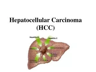
Hepatocellular Carcinoma (HCC). • Different types of cancer can behave very differently . For example , lung cancer and breast cancer are very different diseases . They grow at different rates and respond to

1.43k views • 25 slides

<a href=ACC Cancer Plan" width="320px" />

ACC Cancer Plan

ACC Cancer Plan . Lung Cancer . Best for last ?. F irst for last !. Prevention . Education. Access. ACC Lung Cancer . Education. Prevention . Access. ACC Lung Cancer . Tobacco and Disease: The 5 th Annual Lung Cancer Symposium. November, 2014.

707 views • 56 slides

Non-small-cell lung carcinoma

Non-small-cell lung carcinoma

Non-small-cell lung carcinoma. Non-small-cell lung carcinoma ( NSCLC ) is any type of epithelial lung cancer other than small cell lung carcinoma ( SCLC). As a class, NSCLCs are relatively insensitive to chemotherapy, compared to small cell carcinoma.

758 views • 21 slides

Lung Cancer

Lung Cancer

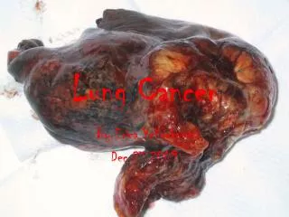
Lung Cancer . By Erica Yellowknee Dec 27 2009. What cause lung cancer?. Smoking, passive smoking, asbestos fibbers, radon gas, familial predisposition, lung diseases, and air pollution cause lung cancer. The main causes is smoking. . Effects of lung cancer .

396 views • 7 slides

Tha role of FDG-PET for Mediastinal Staging in Primary Lung Cancer

Tha role of FDG-PET for Mediastinal Staging in Primary Lung Cancer

Tha role of FDG-PET for Mediastinal Staging in Primary Lung Cancer. Osman Eroğlu, Sedat Ziyade, Alper Toker, Serhan Tanju , Sukru Dilege, Goksel Kalayc ı Istanbul Universty Istanbul Medical School Department of Thoracic Surgery. Mediastinal Evaluation in Lung Cancer. Non-invasive CT

444 views • 17 slides

Lung Cancer

Lung Cancer

Lung Cancer. By: Priyanka , Sonya, and Akshatha. Lung cancer is a cancer that forms in the tissues of the lung. Mostly in the cells lining the air passages. History and Origin. There are two types of cancer: Small cell lung cancer and non-small cell lung cancer

597 views • 10 slides

Mount Vernon Cancer <a href=Network Primary Care Cancer Education Programme" width="320px" />

Mount Vernon Cancer Network Primary Care Cancer Education Programme

Mount Vernon Cancer Network. Wednesday 29 th February 2012 Novotel, Stevenage. Mount Vernon Cancer Network Primary Care Cancer Education Programme. Recognising the Early Signs and Symptoms of Cancers Programme 1: Bowel, Ovarian, Oesophagogastric and Lung Cancers. Mount Vernon Cancer Network.

1.45k views • 127 slides

Mount Vernon Cancer <a href=Network Primary Care Cancer Education Programme" width="320px" />

Mount Vernon Cancer Network Primary Care Cancer Education Programme

Mount Vernon Cancer Network. Wednesday 21 st March 2012 Niland Centre, Bushey. Mount Vernon Cancer Network Primary Care Cancer Education Programme. Recognising the Early Signs and Symptoms of Cancers Programme 1: Bowel, Ovarian, Oesophagogastric and Lung Cancers. Mount Vernon Cancer Network.

1.57k views • 137 slides

Clinical Impression: BRONCHOGENIC CARCINOMA

Clinical Impression: BRONCHOGENIC CARCINOMA

Clinical Impression: BRONCHOGENIC CARCINOMA. Histopathologic image of small cell carcinoma of the lung. CT-guided core needle biopsy. H & E stain. Non-small cell lung carcinoma. 75% of lung cancer Grows and spreads more slowly than small cell Ca

225 views • 11 slides

Lung Cancer

Lung Cancer

Lung Cancer. By: Ashish , Andy, and Megan. Lung Cancer. What is Lung Cancer ? Lung Cancer is the most common type of cancer. Origins of Lung Cancer. What is the History/Origin of Lung Cancer ? Nearly nonexistent in the early 1900’s

796 views • 17 slides

Limited-Stage Small Cell Lung Carcinoma: Overview with Focus on Management

Limited-Stage Small Cell Lung Carcinoma: Overview with Focus on Management

Limited-Stage Small Cell Lung Carcinoma: Overview with Focus on Management. John M. Watkins, M.D. Medical University of South Carolina Department of Radiation Oncology. Background. Small Cell Lung Cancer 15-20% of primary lung neoplasms Decreasing incidence Classically large, central mass

949 views • 69 slides

Clinical Impression: BRONCHOGENIC CARCINOMA

Clinical Impression: BRONCHOGENIC CARCINOMA

Clinical Impression: BRONCHOGENIC CARCINOMA. Histopathologic image of small cell carcinoma of the lung. CT-guided core needle biopsy. H & E stain. Non-small cell lung carcinoma. 75% of lung cancer Grows and spreads more slowly than small cell Ca Types: Adenocarcinoma Squamous cell ca

332 views • 12 slides

Sequential vs. concurrent chemoradiotherapy for locally advanced non-small cell carcinoma

Sequential vs. concurrent chemoradiotherapy for locally advanced non-small cell carcinoma

Sequential vs. concurrent chemoradiotherapy for locally advanced non-small cell carcinoma. Background. Lung cancer remains leading cause of cancer death in UK 5 year survival 8% (all stages) Non-small cell lung cancer accounts for 80% of cases

311 views • 15 slides

Lung Cancer Medical Tourism in Germany

Lung Cancer Medical Tourism in Germany

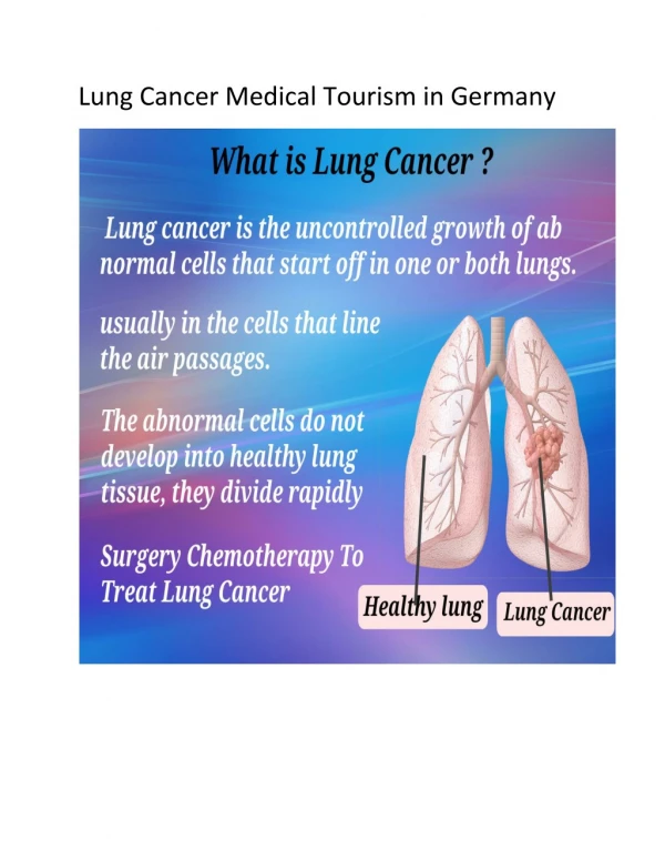
Compass Medical provides Lung Cancer Treatment and Medical Tourism in Germany, Lung Cancer Surgery, Small Cell Lung Cancer and all types Lung Cancer Diagnosis.

146 views • 10 slides

CARCINOMA PANCREAS

CARCINOMA PANCREAS

CARCINOMA PANCREAS. EXOCRINE PANCREASE 5 TH MOST FREQUENT CAUSE OF CANCER DEATH PRECEDED BY LUNG,COLON,BREAST PROSTATE CANCER. CARCINOMA PANCREAS. 1.EPIDEMIOLOGY 2. PATHOGENESIS INCLUDING MOLECULAR LEVEL 3.PATHOLOGY GROSS MICROSCOPY 4. DIAGNOSIS 5. CLINICAL FEATURES

523 views • 45 slides

DNA diagnosis of lung cancer Patrick Willems GENDIA Antwerp, Belgium

DNA diagnosis of lung cancer Patrick Willems GENDIA Antwerp, Belgium

DNA diagnosis of lung cancer Patrick Willems GENDIA Antwerp, Belgium. Treatment of Lung Cancer. Small Cell Lung Cancer (SCLC) chemotherapy radiation Non-Small Cell Lung Cancer (NSCLC) surgery radiation chemotherapy targeted treatment immunotherapy.

723 views • 69 slides